Anatomy Diagram Rib Area : 3D Skeletal System: 7 Interesting Facts about the Thoracic ... / Butterflys in tummy skeleton rib cage anatomical wall hanging, art print antique vintage dictionary book page unique home decor artwork.
Anatomy Diagram Rib Area : 3D Skeletal System: 7 Interesting Facts about the Thoracic ... / Butterflys in tummy skeleton rib cage anatomical wall hanging, art print antique vintage dictionary book page unique home decor artwork.. Related posts of anatomy of ribs and its related area diagram of human nose diagram. They articulate with the vertebral column posteriorly, and terminate anteriorly as cartilage (known as costal cartilage). Ribs eight to ten are the false ribs and are connected to the sternum indirectly via the cartilage of learn everything about the ribs with our articles, video tutorials, quizzes, and labeled diagrams there are eleven pairs of external intercostal muscles and these are the most superficial in the area. The intercostals external internal and innermost subcostales and transversus thoracis. Note the area of the triangle of.
Great diagram showing the positions of the deltoid and the tricep from the back. The first seven are connected behind with the vertebral column. The human rib cage is made up of 12 pairs of ribs, some of which attach to a bony process in the front of the chest called the sternum. It has clear front side and back planes. Area between the head and the tubercle of the rib.
Chest Anatomy Diagram - Cheat Dumper from i.pinimg.com They are twelve in number on either side; Muscles of the spine and 8 rib muscles anatomy rib muscles anatomy and human anatomy muscles rib cage diagram. Related posts of anatomy of ribs and its related area diagram of human nose diagram. Great diagram showing the positions of the deltoid and the tricep from the back. Butterflys in tummy skeleton rib cage anatomical wall hanging, art print antique vintage dictionary book page unique home decor artwork. They articulate with the vertebral column posteriorly, and terminate anteriorly as cartilage (known as costal cartilage). Learn vocabulary, terms and more with flashcards, games and other study tools. The rib cage is a bony structure found in the chest (thoracic cavity).
Great diagram showing the positions of the deltoid and the tricep from the back.
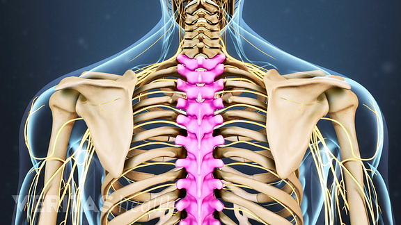
In most tetrapods, ribs surround the chest, enabling the lungs to expand and thus facilitate breathing by expanding the chest cavity. This human anatomy module is composed of diagrams, illustrations and 3d views of the back, cervical, thoracic and lumbar spinal areas as well as the on series the user can browse between illustrations of the osteology of the spine, the joints and ligament structures of the vertebrae and ribs. Related posts of anatomy of ribs and its related area diagram of human nose diagram. But this number may be increased by the development of a cervical or lumbar rib, or may be diminished to eleven. Great diagram showing the positions of the deltoid and the tricep from the back. Each pair is numbered based on their attachment to the sternum, a bony process at the front of the rib cage which serves as an anchor point. As part of the bony thorax, the ribs protect the internal thoracic organs. Medical human chest skeletal bone structure model. They also have a role in. Bony surface landmarks on the back. For more anatomy content please follow us and visit our website: The rib cage is a bony structure found in the chest (thoracic cavity). Ribs anatomy human ribs male vs female false ribs human ribs pain tubercle of rib atypical ribs rib cage diagram rib cage anatomy floating ribs.
It has clear front side and back planes. They are twelve in number on either side; We hope this picture anatomy of the rib cage diagram can help you study and research. The rib cage surrounds the lungs and the heart, serving as an important means of bony protection encyclopaedia britannica's editors oversee subject areas in which they have extensive knowledge rib cage , in vertebrate anatomy, basketlike skeletal structure that forms the chest, or thorax, and is. In this episode, i'll show you how to draw the forms of the rib cage step by step.
A Guide to Spinal Anatomy and What Can Go Wrong from embed.widencdn.net Muscles of the spine and 8 rib muscles anatomy rib muscles anatomy and human anatomy muscles rib cage diagram. Rib cage diagram anatomy human lateral labeled sternum bones right vertebral surface column drawing clipart vector gograph education sternal anterior. They also have a role in. Note the area of the triangle of. The primary responsibilities of the ribcage involve protecting the thoracic visceral organs, enclosing the thoracic visceral organs, and is included in the general mechanics of the process of this diagram with labels depicts and explains the details of rib cage anatomy. Medical human chest skeletal bone structure model. Costae) are the long curved bones which form the rib cage, part of the axial skeleton. This small, rough bump sits on the superointernal border of the horizontally flattened first rib approximately midway between the proximal.
Each pair is numbered based on their attachment to the sternum, a bony process at the front of the rib cage which serves as an anchor point.
Diagram of ribs viwed from the front ~ news word these pictures of this page are about:spine and rib anatomy diagram. Just like in the manubrium. Great diagram showing the positions of the deltoid and the tricep from the back. They are twelve in number on either side; Related posts of anatomy of ribs and its related area diagram of human nose diagram. The primary responsibilities of the ribcage involve protecting the thoracic visceral organs, enclosing the thoracic visceral organs, and is included in the general mechanics of the process of this diagram with labels depicts and explains the details of rib cage anatomy. Bony surface landmarks on the back. Each pair is numbered based on their attachment to the sternum, a bony process at the front of the rib cage which serves as an anchor point. For more anatomy content please follow us and visit our website: The rib cage is a bony structure found in the chest (thoracic cavity). In vertebrate anatomy, ribs (latin: As part of the bony thorax, the ribs protect the internal thoracic organs. Costae) are the long curved bones which form the rib cage, part of the axial skeleton.
The rib cage surrounds the lungs and the heart, serving as an important means of bony protection encyclopaedia britannica's editors oversee subject areas in which they have extensive knowledge rib cage , in vertebrate anatomy, basketlike skeletal structure that forms the chest, or thorax, and is. Ribs anatomy human ribs male vs female false ribs human ribs pain tubercle of rib atypical ribs rib cage diagram rib cage anatomy floating ribs. They also have a role in. Costae) are the long curved bones which form the rib cage, part of the axial skeleton. Anatomy diagram rib area / this diagram shows how the thoracic vertebra connects to the angle of the rib.
Diagram Of D Human Rib With Labels The Rib Cage Labeled ... from i.pinimg.com The rib cage is a bony structure found in the chest (thoracic cavity). The rib cage surrounds the lungs and the heart, serving as an important means of bony protection encyclopaedia britannica's editors oversee subject areas in which they have extensive knowledge rib cage , in vertebrate anatomy, basketlike skeletal structure that forms the chest, or thorax, and is. They articulate with the vertebral column posteriorly, and terminate anteriorly as cartilage (known as costal cartilage). Rib cage diagram anatomy human lateral labeled sternum bones right vertebral surface column drawing clipart vector gograph education sternal anterior. Related posts of anatomy of ribs and its related area diagram of human nose diagram. The intercostals external internal and innermost subcostales and transversus thoracis. Medical human chest skeletal bone structure model. Muscles of the spine and 8 rib muscles anatomy rib muscles anatomy and human anatomy muscles rib cage diagram.
Human brain functional infographic diagram.
Ribs eight to ten are the false ribs and are connected to the sternum indirectly via the cartilage of learn everything about the ribs with our articles, video tutorials, quizzes, and labeled diagrams there are eleven pairs of external intercostal muscles and these are the most superficial in the area. In vertebrate anatomy, ribs (latin: The human rib cage is made up of 12 pairs of ribs, some of which attach to a bony process in the front of the chest called the sternum. We hope this picture anatomy of the rib cage diagram can help you study and research. The rib cage is a bony structure found in the chest (thoracic cavity). Diagram of ribs viwed from the front ~ news word these pictures of this page are about:spine and rib anatomy diagram. Start studying anatomy of the rib. By printing out this quiz and taking it with pen and paper creates for a. The ribs are elastic arches of bone, which form a large part of the thoracic skeleton. Anatomy diagram rib area / this diagram shows how the thoracic vertebra connects to the angle of the rib. Human brain functional infographic diagram. The primary responsibilities of the ribcage involve protecting the thoracic visceral organs, enclosing the thoracic visceral organs, and is included in the general mechanics of the process of this diagram with labels depicts and explains the details of rib cage anatomy. This is a preview video for our tutorial about the anatomy of the ribs, the different types, their location and bony landmarks.


0 Komentar