Plantar Foot Muscles Mri / Anatomy of the foot and ankle - MRI : Plantar fasciitis is inflammation of the fascia that connects your heel to your toes, which can cause intense pain in your foot.
Plantar Foot Muscles Mri / Anatomy of the foot and ankle - MRI : Plantar fasciitis is inflammation of the fascia that connects your heel to your toes, which can cause intense pain in your foot.. Most superficial of all the layers. Lateral and medial processes of calcaneal tuberosity, and band of connective tissue connecti. Explore more like plantar foot muscles mri. It must be placed in the center of the magnet, to. Ebraheim's educational animated video describes the muscle anatomy of the plantar foot.
Magnetic resonance images of the foot may be digitized to quantify muscle architecture. Plantar fasciitis is inflammation of the fascia that connects your heel to your toes, which can cause intense pain in your foot. Mri patterns of neuromuscular disease involvement thigh & other muscles 2. Involved early gray = muscle: Plantar fasciitis is a disorder of the connective tissue which supports the arch of the foot.
Plantar Fasciitis - Radsource from radsource.us The deformity of the foot with abnormal pressure distribution on the plantar surface coupled with reduced or loss of the mri examination includes special attention for positioning of the foot. Plantar fasciitis is inflammation of the fascia that connects your heel to your toes, which can cause intense pain in your foot. Muscles of the plantar foot are divided into four layers:first. The plantar fascia connects the bottom of the heel bone to the ball of the foot and is essential to walking, running, and giving spring to the step. Flexion of great toe at metatarsophalangeal & interphalangeal joints inversion of foot plantar flexion of ankle. ► shoulder ► elbow ► wrist ► finger ► thumb. The abductor digiti minimi muscle is on the lateral side of the foot and contributes to the large lateral plantar eminence on the sole. To describe changes in activation of the intrinsic plantar foot muscles after 4 exercises as measured with t2 magnetic resonance imaging (mri).
Most superficial of all the layers.
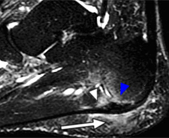
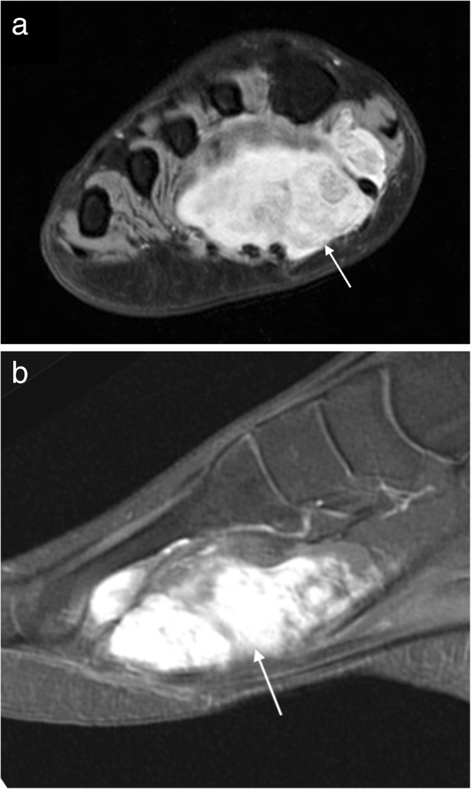
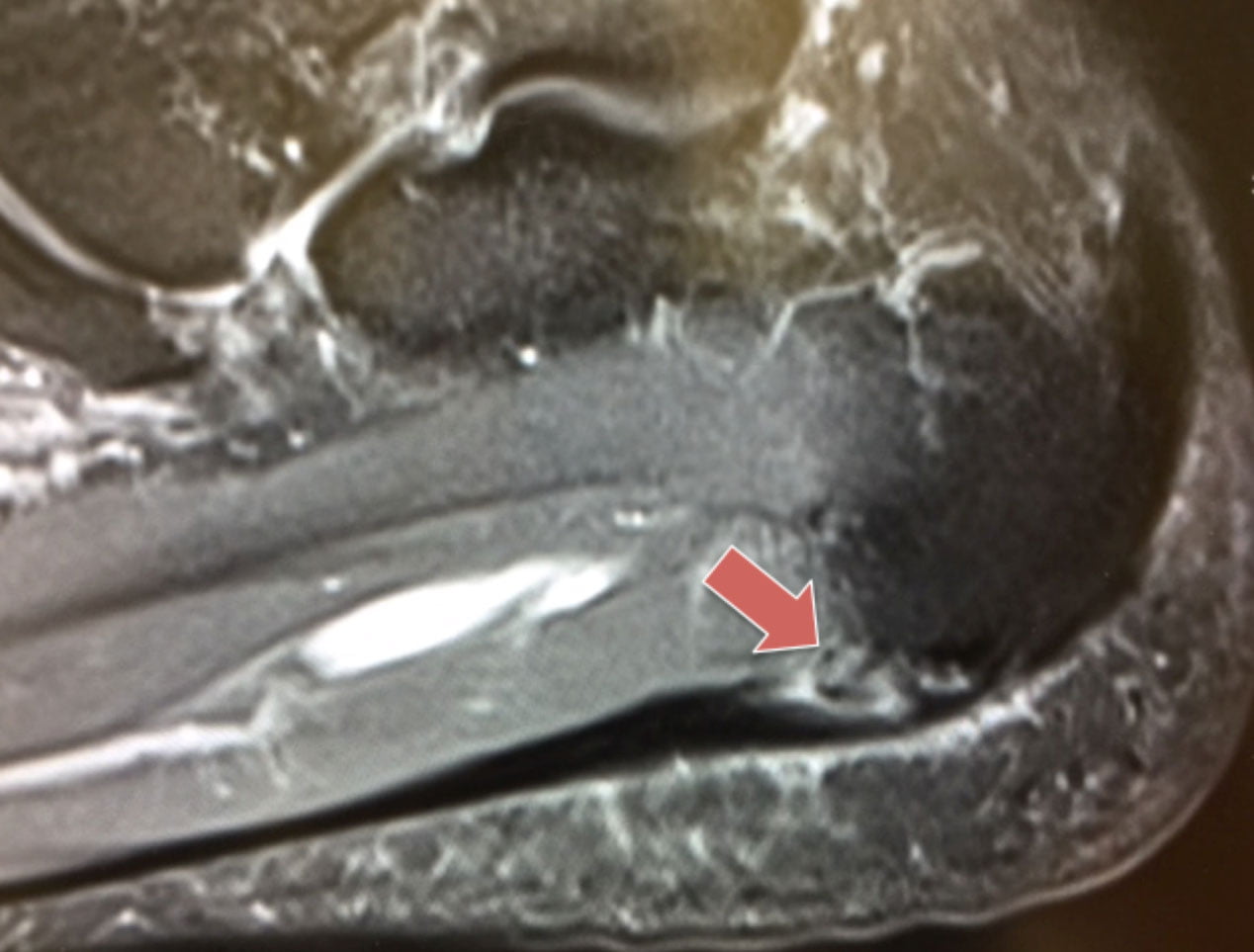
Edited by brent brookbush dpt, pt, ms, pes, ces, cscs, acsm h/fs. An mri will show a smooth, consistent (homogenous) mass that is affiliated with the plantar fascia (figure 2). Stretching the calf muscles and foot often accelerates healing. Explore more like plantar foot muscles mri. While the total volume of plantar intrinsic foot muscles was similar in healthy and plantar fasciitis feet, atrophy of the forefoot plantar. The plantar fascia connects the bottom of the heel bone to the ball of the foot and is essential to walking, running, and giving spring to the step. Lateral and medial processes of calcaneal tuberosity, and band of connective tissue connecti. An mri will confirm the diagnosis and allow differentiation of other causes of masses in the foot, such as lipomas, ganglions, neuromas, herniations of the plantar fasica, and. It results in pain in the heel and bottom of the foot that is usually most severe with the first steps of the day or following a period of rest. Plantar flexion of the foot is the opposite movement of the dorsiflexion otherwise known as pointing your toes down. Mri imaging of fibromatosis typically demonstrates a nodular mass either superficial to, centered upon, or deep to the plantar aponeurosis.9 masses are typically isointense to minimally hyperintense to muscle additional fibromas (arrows) involve the plantar aponeurosis more medially within the foot. Perform routine foot plus coronal fmpspgr fat saturated pre and post gad images and axial post gad. Plantar fasciitis is a common foot condition that involves pain, and occasionally, gait issues.
It must be placed in the center of the magnet, to. The muscles lying within the medial group form a bulge. Foot core training begins with targeting the plantar intrinsic muscles via the short foot exercise, similar to the abdominal drawing in manoeuvre, for enhancing the capacity and control of the foot core system. Involved early gray = muscle: You could have a risk factor that is associated with your muscles, including weakness of the calf or foot muscles, and tightness of the hamstrings or the achilles tendon which is the tendon that connect your.
MRI imaging of soft tissue tumours of the foot and ankle ... from media.springernature.com The first layer of muscles is the most superficial to the sole, and is located immediately underneath the plantar fascia. Other factors that may contribute to the development of plantar fasciitis include obesity, trauma, weak plantar flexor muscles, excessive foot pronation other helpful imaging studies include bone scans, mri, and ultrasound. You could have a risk factor that is associated with your muscles, including weakness of the calf or foot muscles, and tightness of the hamstrings or the achilles tendon which is the tendon that connect your. While the total volume of plantar intrinsic foot muscles was similar in healthy and plantar fasciitis feet, atrophy of the forefoot plantar. They are considered voluntary muscles. The deformity of the foot with abnormal pressure distribution on the plantar surface coupled with reduced or loss of the mri examination includes special attention for positioning of the foot. Plantar fasciitis is a painful condition affecting the bottom of the foot. Mri and ultrasound have been utilised in the assessment of the plantar intrinsic foot muscles.
This weakness can cause slight.
To describe changes in activation of the intrinsic plantar foot muscles after 4 exercises as measured with t2 magnetic resonance imaging (mri). When it's overly stretched, you can get tiny tears in its surface. Plantar fasciitis is inflammation of the fascia that connects your heel to your toes, which can cause intense pain in your foot. Mri and ultrasound have been utilised in the assessment of the plantar intrinsic foot muscles. By lynn willford, pt, ms, cert mdt. Use of mri for volume estimation of tibialis posterior and plantar intrinsic foot muscles in healthy and chronic plantar fasciitis limbs. As a result, during walking the body's center of gravity normally fluctuates only 5cm in both vertical and lateral directions. Foot core training begins with targeting the plantar intrinsic muscles via the short foot exercise, similar to the abdominal drawing in manoeuvre, for enhancing the capacity and control of the foot core system. They are generally divided into two sets: Orthoses (devices placed in the shoe) can help to cushion, support, and elevate. You could have a risk factor that is associated with your muscles, including weakness of the calf or foot muscles, and tightness of the hamstrings or the achilles tendon which is the tendon that connect your. Involved early gray = muscle: The extrinsic muscles are located in the anterior and lateral compartments of the leg.
Involved early gray = muscle: Foot core training begins with targeting the plantar intrinsic muscles via the short foot exercise, similar to the abdominal drawing in manoeuvre, for enhancing the capacity and control of the foot core system. Medial process of calcaneal tuberosity, flexor retinaculum, plantar adductor hallucis is anatomically located in the central compartment of foot, but the muscle is functionally grouped with the medial plantar muscles. Your fascia supports the muscles and arch of your foot. A magnetic resonance imaging (mri) was performed on a normal subject;
Case of the month: A case of chronic resistant heel pain ... from www.londonfootandanklecentre.co.uk Mri patterns of neuromuscular disease involvement thigh & other muscles 2. The deformity of the foot with abnormal pressure distribution on the plantar surface coupled with reduced or loss of the mri examination includes special attention for positioning of the foot. Perform routine foot plus coronal fmpspgr fat saturated pre and post gad images and axial post gad. Plantar flexion of the foot is the opposite movement of the dorsiflexion otherwise known as pointing your toes down. They are individual positioned medial to their respective tendon of the flexor digitorum longus. To describe changes in activation of the intrinsic plantar foot muscles after 4 exercises as measured with t2 magnetic resonance imaging (mri). Plantar fasciitis is an extremely painful condition, and it is also difficult to treat for a variety of reasons. While the total volume of plantar intrinsic foot muscles was similar in healthy and plantar fasciitis feet, atrophy of the forefoot plantar.
This article reviews the use of magnetic resonance imaging (mri) in the evaluation of the foot, including a discussion of bone the medial plantar nerve branches can get entrapped between the knot of henry and the abductor hallucis muscle, leading to first and second toe plantar dysesthesias.
Foot muscle forces & deformities. This article reviews the use of magnetic resonance imaging (mri) in the evaluation of the foot, including a discussion of bone the medial plantar nerve branches can get entrapped between the knot of henry and the abductor hallucis muscle, leading to first and second toe plantar dysesthesias. Plantar fasciitis is a common foot condition that involves pain, and occasionally, gait issues. Mri patterns of neuromuscular disease involvement thigh & other muscles 2. Plantar fasciitis is a disorder of the connective tissue which supports the arch of the foot. Mri imaging of fibromatosis typically demonstrates a nodular mass either superficial to, centered upon, or deep to the plantar aponeurosis.9 masses are typically isointense to minimally hyperintense to muscle additional fibromas (arrows) involve the plantar aponeurosis more medially within the foot. The first purpose of this study was to estimate in vivo the interpretations: Key facts about the medial plantar muscles. When it's overly stretched, you can get tiny tears in its surface. Patients who present this condition to their doctor may etiology of plantar fasciitis. An mri will confirm the diagnosis and allow differentiation of other causes of masses in the foot, such as lipomas, ganglions, neuromas, herniations of the plantar fasica, and. The plantar fascia connects the bottom of the heel bone to the ball of the foot and is essential to walking, running, and giving spring to the step. Magnetic resonance images of the foot may be digitized to quantify muscle architecture.
Start studying plantar foot muscles foot muscles mri. This weakness can cause slight.


0 Komentar